CERVİCAL HERNİATED DİSCS
Cervical Herniated Disc Surgeries
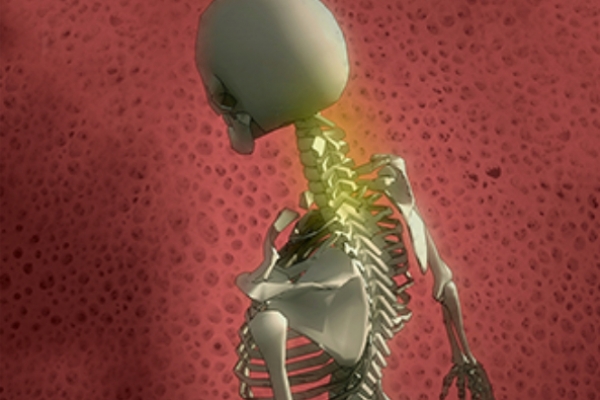
Although cervical disc hernias may cause severer damages than lumbar herniated discs when they grow, due to their locations, they are more likely to melt away without treatment. The spinal cord begins at the end of the brain stem, and it transmits electrical impulses coming from the brain along the 7 cervical vertebrae to the cervical region (the neck), the thoracic region (the back) and the lumbar region (the waist), respectively.
This disease encountered by mostly people who are engaged in desk jobs, such as teachers, secretaries, and those who sit too much at the computer, manifest itself as a pain in the nape of the neck, a headache, a pain affecting the both scapulae, and a pain extending up to the shoulder and elbow. In advanced cases, dizziness and imbalance appear, and these symptoms are followed by urinary and fecal incontinence as well as sexual dysfunctions (impotence) in men.
Cervical herniated disc pains usually aggravates at night. It significantly reduces the sleep quality of the patient. When the pain is felt in the shoulder and arm, the patient try to relieve the pain by holding his/her arm on the head or the nape of the neck.
Most of admissions to polyclinics due to neck pain involve the cases of flattened necks (increased cervical lordosis) or cervical vertebrae deformities (kyphosis, scoliosis). In the treatment of such conditions encountered due to weakness in the cervical muscles, misuse of the neck, and vitamin deficiencies (B12) usually involves the use of painkillers; and with the alleviation of the pain, patients are instructed to do strengthening physiotherapy exercises, with intent to ensure a permanent pain relief.
Surgery is needed for patients diagnosed with an advanced loss of strength, muscular atrophy (atrophy), or a scar (myelomaleous) on their spinal cord detected in a cervical MRI scan.
Cervical herniated disc surgeries are performed under a microscope as well; and they are called cervical microdiscectomy.
The surgery is performed with a 2-3 cm incision on the front side of the neck, extending from the midline to the side. A right handed surgeon opens the site from the right hand side, while a left handed surgeon performs the operation by working from the left hand side.
Since cervical herniated disc surgeries are performed from the front side rather than the back side as in lumbar herniated disc surgeries, the whole cartilaginous tissue and herniated material are removed, and then disc implants are placed in their places, which function as a material fixing the joint in its place and protecting the structure and cartilage movements. No hernia forms in the operated site, i.e. cervical herniated discs recur. The surgery takes less than 1 hour. By accessing through the front side of the neck, the carotid artery and muscles are pushed outwardly, while the trachea-esophagus is pushed inwardly for their exclusion. The joint tissue constituting the joint between two vertebrae is completely removed under a microscope, and then the herniated part that presses the spinal cord is completely cleaned. The wound is stitched from inside, and is covered with sterile strips. Once the patient regains consciousness after surgery, he /she is allowed to walk. The patient does not need to use a neck protector. The patient, who is allowed to walk whenever he/she wants, will not feel the pain that he/she had felt in his/her shoulders and arms before the surgery. He can be discharged from the hospital on the day of the surgery or the next morning. Once the patient arrives home, he/she will take a bath and remove the adhesive tapes from the scar. The patient is asked to go for a checkup after the 1-week rest period that involves no restriction.


